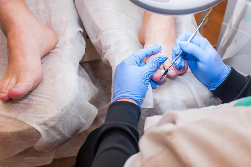
Fungal nail infections -diagnosis and new treatment therapies
M3 India Newsdesk Nov 27, 2018
The incidence of fungal nail infections, (onychomycosis) is fairly common, in warm humid climates such as India. Here is a quick overview of the diagnosis and treatment therapies used for the disease.

Onychomycosis, a common infection of the nail is caused by a variety of fungi- dermatophytes, non-dermatophytes and Candida. Pain, discomfort, and impaired/lost tactile functions are some of the symptoms seen in onychomycosis cases of the fingernails. Besides this, dystrophy of the toenails may lead to difficulty in walking, exercise, and improper fitting shoes. Therefore, both psychosocial and negative physical effects are associated with onychomycosis.
Dermatophytic onychomycosis
The most common etiological fungi that cause onychomycosis in 90% of toenails and 50% of fingernails are dermatophytes. When dermatophytes also invade the nail plate, this condition is known as tinea unguium. Tinea unguium is most frequently caused by Trichophyton rubrum (T. rubrum) followed by T. mentagrophytes as the second most common fungi. Toenails are most commonly affected whereas fingernails are occasionally affected by non-dermatophyte molds (NDM) also.
Diagnosis
- Samples for diagnosing tinea pedis, or manuum should be taken separately from fingernails and toenails after the patients has stopped both topical and systemic antifungal drugs for a minimum of 2-4 weeks.
- The simplest and fastest method for confirming fungal nail infection is by direct microscopy.
- In order to confirm the diagnosis and to ascertain the exact causative fungus, a culture is necessary. Culturing of the nail specimens can be done by incubating specimens using different media at 25 to 30°C.
- Primary medium contains cycloheximide and can detect most NDM and bacteria, e.g. DTM, mycosel (BBL), and mycobiotic (DIFCO).
- Sabouraud glucose agar (SGA), Littman's Oxgall medium, and potato dextrose agar (PDA) are all secondary media which are free of cycloheximide and that allow isolation of NDM.
Note: To negate bacterial contamination, antibiotics such as chloramphenicol and gentamicin can be added to SGA or PDA.
- In cases of suspected onychomycosis, histopathology of nail specimens might become compulsory when the KOH and cultures are repetitively negative.
- Stains which are more selective than PAS include the Grocott Methenamine silver and calcofluor white (CFW) stains.
- Immunohistochemistry and dual flow cytometry are less commonly used tests to diagnose onychomycosis. These tests are mainly used for the identification of mixed infections and for quantifying of the fungal load in the nail.
- Recently DNA-based methods such as PCR-RFLP assays have been utilised for checking for the presence of fungi.
Treatment of dermatophytic onychomycosis
Treatment can be done either by topical methods, systemic methods, or a combination of the two. In a recent study, the novel azole antifungal voriconazole had a lower MIC against dermatophytes, Candida and NDM.
- Ciclopirox 8% and amorolfine 5% lacquers have a broad action towards yeasts, dermatophytes, and NDM and therefore are the commonly used topical antifungals presently.
- Griseofulvin, azoles such as ketoconazole, itraconazole and fluconazole, and the allylamine terbinafine are the oral drugs used for the treatment of onychomycosis.
Newer therapies for dermatophytic onychomycosis
To enhance nail penetration when using topical drugs, novel strategies include:
- Iontophoresis
- Physical methods such as manual and electrical nail abrasion
- Acid etching
- Microporation
- Applying low-frequency ultrasound
- Laser nail ablation
- Use of chemical ungual activity enhancer
Laser treatment
It is the most recent addition in the armamentarium to treat onychomycosis.
- The efficacy of the Noveon dual-wavelength (using 870-nm, 930-nm light) near-infrared diode laser in treating moderate to severe onychomycosis has been seen.
- Promising outcomes are also being seen using a new 0.65 millisecond pulsed Nd:YAG 1064-nm laser and femtosecond infrared titanium sapphire laser.
- The efficacy of topical germicidal UVC radiation has also been shown for the treatment of onychomycosis.
- Mixed success has been seen with photodynamic therapy.
Combination therapy
Not only does combination therapy improve efficacy and reduce relapse, but when both oral and topical drugs are used together, taking lower doses of the oral drug may result in better patient tolerance and compliance. In patients in which it is likely that antifungal therapy may fail, antifungals may be given as combination therapy, either in parallel (both oral and topical drugs given simultaneously) or in a sequential form (administration of oral drug alone, followed by topical).
In sequential therapy, the combined use of two oral antifungals that act on two different pathways in ergosterol metabolism is seen. In order to produce sensitive hyphae that are less refractory to antifungal treatment, boosted oral antifungal treatment (BOAT) can be used to target dormant chlamydospores and arthroconidia within the nail plate.
Surgery
Part removal (debridement) or complete removal (avulsion) of the nail plate can also be achieved by surgical methods. In cases where a patient may have lateral nail plate involvement or onycholytic pockets on the undersurface of the nail filled with a longitudinal spike/dermatophytoma, partial removal is a good alternative.
Treatment of non-dermatophytic onychomycosis
- A surgical avulsion (~60%), when combined with topical antifungals, shows a better cure rate than monotherapy with terbinafine or itraconazole alone (20% Acremonium species, 29% Fusarium, and 42% Scopulariopsis brevicaulis).
- A better response with systemic therapy using terbinifine or itraconazole is seen only in Aspergillus and can be regarded as an exception.
- In cases of superficial white onychomycosis, systemic therapy should be added when it is originating from the proximal nail fold.
- Itraconazole or terbinafine can also be used to treat distal and lateral subungual onychomycosis.
Candida onychomycosis
- Itraconazole and fluconazole have been found to be equally effective in both systemic and topical treatment of candida onychomycosis. Both continuous and pulse regimens of itraconazole can be used.
- The dose recommendations of fluconazole in cases of candida onychomycosis are 50 mg/day or 300 mg/week to be given for a minimum of 4 weeks for fingernail onychomycosis, and 12 weeks for toenail onychomycosis.
- The combination of amorolfine nail lacquer and 2 pulses of itraconazole were found to be safer, cheaper and more effective than monotherapy with 3 pulses of itraconazole in moderate and severe cases of candida onychomycosis.
Treatment of onychomycosis in special populations
Close family contacts and children affected with onychomycosis should undergo careful screening for other superficial mycoses.
Children & Teens
- Griseofulvin at a dose of 10 mg/kg/day is the only approved antifungal for children
- Terbinafine and itraconazole have been used in patients in the age group of 4 to 17 years with no significant children specific side effects
- Fluconazole (3 to 6 mg/kg) is also considered safe in daily doses and in pulsed doses
Elderly patients
- For the elderly, both continuous and pulsed terbinafine and itraconazole are found to be safe
- For dermatophyte onychomycosis, terbinafine is the drug of choice due to its greater mycological cure, lesser side effects, lesser drug interactions, and lower cost when compared to continuous itraconazole therapy
Diabetic patients
Onychomycosis prevalence in diabetic men is 2.5 to 2.8 times more than in the control group population. Yeast is found to be the most common causative agent in this group followed by dermatophytes and NDM.
- Topical therapies in combination with foot care intervention such as nail drilling have been found to be effective
- Due to its low risk of drug interaction, and its good efficacy, terbinafine is regarded as the first line of therapy
This article is part of a series on Fungal Infections. Click on the links below to read the other articles in the series.
Guideline on management of dermatophytosis in India
Managing fungal infections in diabetes: Dr Kiran Godse & Dr. Dakshata Tare
-
Exclusive Write-ups & Webinars by KOLs
-
Daily Quiz by specialty
-
Paid Market Research Surveys
-
Case discussions, News & Journals' summaries