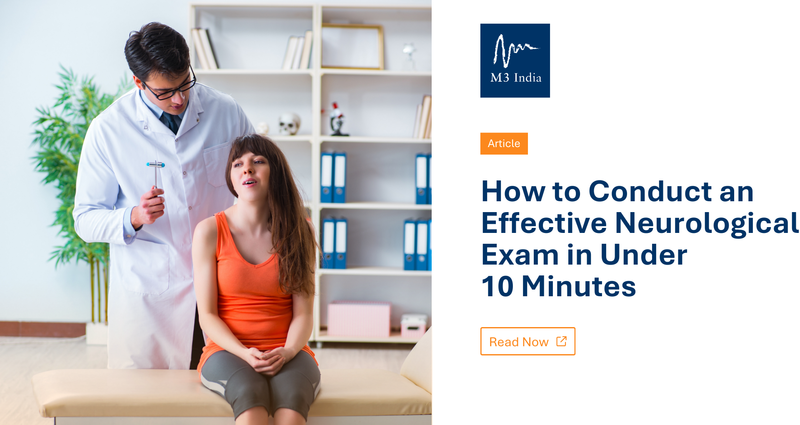
Article: How to Conduct an Effective Neurological Exam in Under 10 Minutes
M3 India Newsdesk Apr 14, 2025
This article provides an organised method for the neurological examination with a focus on lesion localisation by clinical observation and case-based learning,

The neurologic examination is considered by many to be daunting. It may seem tedious, time-consuming, overly detailed, idiosyncratic, and even capricious. Every neurologist has his/her own version of the examination and may appear to use “magical thinking” to come up with a diagnosis at the end.
In reality, the examination is quite simple. When performing the neurological examination, it is important to keep the purpose of the examination in mind, namely, to localise the lesion. A basic knowledge of neuroanatomy is necessary to interpret the examination.
The key to performing an efficient neurological examination is observation. More than half of the neurological examination is performed by simply observing the patient – how he/she speaks, thinks, walks, moves, and interacts with the examiner. A skillful observer will be able to localise a lesion based on simple observations. Formalised testing merely refines the diagnosis and may only require several additional steps.
Performing an overly detailed neurological examination without a purpose in mind is a waste of time and often yields incidental findings that cloud the picture.
Components of the 10-minute Neurological Examination
1. Mental Status
- Cognition is essentially tested during history taking.
- Language is also tested during history taking, except for naming.
2. Cranial Nerves
- Don’t forget visual fields by confrontation – vision is processed by 1/3 of the cerebral hemispheres.
- Check Pupils and Eye Movements – don’t forget to test saccades as well as pursuits
- Facial strength is best tested by observing the patient for asymmetries during natural speech; also observe for symmetry of eye blinks.
- Lower cranial nerves (IX-XII) only need to be tested if dysphagia and dysarthria are present.
3. Motor Examination
- Adventitial Movements – tics, tremor and bradykinesia are best observed during history taking
- Pronator Drift – implies upper motor neuron dysfunction
- External Rotation of Leg – implies upper motor neuron dysfunction
- Muscle Tone – key examination point – important for diagnosing subtle upper motor neuron lesions and Parkinson’s disease
- Functional Strength testing – more important than formal push-pull testing, more reliable, and quicker!
4. Sensory Examination
- Focus on sensory testing to the patient’s symptoms.
- Sensory testing is purely subjective, so don’t over-interpret.
- Check for sensory level on the back if a spinal cord lesion is suspected.
- Touching nose with eyes closed – an excellent test of proprioception.
- The Romberg test tests proprioception (peripheral nerves and dorsal columns) and is not a test of cerebellar function.
5. Coordination
- Many things cause ataxia – cerebellar lesions, sensory disorders and upper motor neuron lesions.
- Don’t forget truncal stability – truncal ataxia implies a lesion of the cerebellar vermis.
6. Reflexes
- The only purely objective part of the neurological exam.
- Look for asymmetries and sustained clonus.
- Don’t over-interpret the Babinski sign.
7. Gait
- Perhaps the most important part of the 5-minute neurological exam.
- Look at the base, stride, arm-swing, turns and symmetry.
Order of the 10-minute Neurological Examination
1. Mental status, adventitial movements and facial symmetry (already tested during history taking)
2. Gait (casual, heel, toe, tandem)
3. Truncal stability (vermis) and Romberg test (proprioception)
4. Functional motor testing
- Lower limbs - arise from a squat (or a chair with arms folded)
- Upper limbs - raise arms above head
5. Visual fields, pupils and eye movements
6. Motor exam
- Pronator drift
- Finger-to-nose testing with eyes closed
- Motor tone d. Hand grips
7. Sensory exam (already performed with Romberg and finger-to-nose testing)
8. Coordination (already performed with truncal stability and finger-to-nose testing)
9. Reflexes
- Muscle stretch reflexes
- Babinski sign
Neurological Diagnosis
The Neurological history and physical examination are the most important tools in neurological diagnosis. Although confirmatory laboratory data, including modern imaging techniques such as CT scanning and magnetic resonance imaging, have provided further accuracy in neurologic diagnosis, the history and physical examination remain the mainstays.
Neurological diagnosis can be divided into two types: anatomic and aetiologic.
The Anatomic Diagnosis
It localises the lesion within a specific area of the neuraxis, i.e. cerebral hemispheres, diencephalon, brain stem, spinal cord, or the peripheral nervous system. Findings on neurologic examination are most important in making an anatomic diagnosis.
The Etiologic Diagnosis
It specifies the cause of the lesion and is mainly obtained from information provided by the neurologic history. The time course of the illness often helps define the etiologic agent responsible for causing the anatomic lesion. Several examples follow:
- Lesions of Sudden Onset are typically due to vascular accidents, such as stroke.
- Slowly Progressive Lesions are typically due to expanding mass lesions, such as a tumor or abscess.
- Lesions with Exacerbating and Remitting Courses are frequently due to demyelination, such as can be seen with multiple sclerosis.
- Relentlessly Progressive Lesions Involving Diffuse Areas of the Nervous System are typically due to nutritional deficits or to degenerative disorders of the brain and nervous system.
Case Studies
Case 1
A 68-year-old woman with hypertension was brought to the emergency department by her friend because of dizziness, vertigo and difficulty walking, which she first noted when she awoke from a nap that evening.
BP=200/130 mm Hg and P=76/min. She is examined lying on a gurney in the emergency department. There is minimal nystagmus with right gaze. Facial strength and sensation are normal. Motor and sensory examinations are entirely normal. Finger-to-nose testing is normal bilaterally. Muscle stretch reflexes are normal throughout.
Questions
- Where would you best localise the lesion?
- What is the most likely Diagnosis?
- What is the most appropriate next step in diagnosis?
Next Step
- Walk the patient
- Significant truncal ataxia
Diagnosis
Cerebellar Hemorrhage
Case 2
A 55-year-old man with hypertension and diabetes mellitus is admitted for cardiac catheterisation because of worsening angina and an abnormal exercise tolerance test. Following the procedure, which demonstrates severe LAD disease, he is noted to be confused, and a neurological consultation is obtained.
BP=150/90 mm Hg, and P=80/min and regular. He is awake, alert and fully oriented. He appears confused when asked to describe what happened to him that day. His face is symmetrical. He has full power in all four limbs. Sensory examination is entirely normal. Muscle stretch reflexes are symmetrical.
Questions
- Where would you best localise the lesion?
- What is the most likely diagnosis?
- What is the most appropriate next step in diagnosis?
Next Step
- Test naming -Significant anomia
- Test visual fields -Right homonymous hemianopia
Diagnosis
Left MCA embolic stroke
Case 3
A 60-year-old college professor is referred for evaluation of muscle stiffness and a weak voice that has been present for the past six months. Some of his students have complained that it is getting more difficult to understand him when he lectures. They also have had trouble reading his handwriting on the blackboard.
BP=160/80 mm Hg and P=68/min. Mental status is normal. His voice is soft, but cranial nerves are otherwise normal. Muscle power is full, and sensation is normal in all four limbs. Muscle stretch reflexes are 2+ throughout and plantar responses are flexor bilaterally. His gait is somewhat slow but is otherwise normal.
Questions
- Where would you best localise the lesion?
- What is the most likely diagnosis?
- What is the most appropriate next step in diagnosis?
Next Steps
- Look for extra-pyramidal signs: Cogwheel rigidity at the wrists, R>L
- Bradykinesia: slowed finger taps, R>L
- Intermittent resting tremor at his right wrist
- Postural instability
Diagnosis
Parkinson disease
Disclaimer: The views and opinions expressed in this article are those of the author and do not necessarily reflect the official policy or position of M3 India.
About the author of this article: Dr Nikhil Repaka is a neuro-physician at Jagruth Super Specialty Hospital, Khammam.
-
Exclusive Write-ups & Webinars by KOLs
-
Daily Quiz by specialty
-
Paid Market Research Surveys
-
Case discussions, News & Journals' summaries