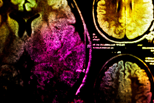
Multicolour MRIs could improve disease diagnosis
IANS Aug 17, 2017
Researchers have developed a method that could make magnetic resonance imaging - MRI - multicolour and help improve disease diagnosis.

Current MRI techniques rely on a single contrast agent injected into a patient's veins to vivify images.The new method uses two at once, which could allow doctors to map multiple characteristics of a patient's internal organs in a single MRI. The strategy could serve as a research tool and even aid disease diagnosis."The method we developed enables, for the first time, the simultaneous detection of two different MRI contrast agents," said Chris Flask, Associate Professor at Case Western Reserve University School of Medicine in Cleveland, Ohio, US. Two contrast agents could include one specifically targetting diseased tissue, and one designed to show healthy tissue, for example. The new method would enable immediate comparisons of how each agent distributes in the same patient.
"This multi-agent detection capability has the potential to transform molecular imaging, as it provides a critical translational pathway for studies in patients," said Flask.In a study published in the journal Scientific Reports, the researchers described how two contrast agents, gadolinium and manganese, can be detected and independently quantified during MRIs. The results provide "an adaptable, quantitative imaging framework to assess two MRI contrast agents simultaneously for a wide variety of imaging applications", the researchers said."In this initial paper, we validated our new methodology, opening the possibility for numerous follow-on application studies in cancer, genetic diseases such as cystic fibrosis, and metabolic diseases such as diabetes," Flask said.
-
Exclusive Write-ups & Webinars by KOLs
-
Daily Quiz by specialty
-
Paid Market Research Surveys
-
Case discussions, News & Journals' summaries