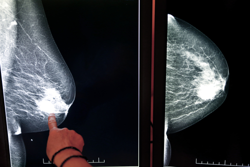
Fluorescence imaging to identify cancer-affected breast tissues introduced in AIIMS
PTI Jul 29, 2019
In a first, a state-of-the-art fluorescence imaging technology for easy identification of cancer-affected tissues in breast has recently been introduced at the AIIMS here.

According to Dr SVS Deo, Head of the Department of Surgical Oncology at AIIMS, the technology will be a "game changer" in breast cancer surgery space as it precisely helps identify relevant tissue intraoperatively.During breast cancer surgery, surgeons inject a safe and affordable indocyanine green (ICG) dye in patients. Using Fluorescence Imaging technology, surgeons can view blood flow in vessels, micro-vessels, tissue perfusion and critical anatomical structures intraoperatively.
"The relevant tissues light up in fluorescent green colour. The reliability and multiple applications of the imaging are a significant differentiation compared to currently used technologies like blue dye," he said."Due to lack of this critical information, earlier all lymph nodes including healthy ones would be removed completely causing significant collateral damage to patient. With Fluorescence Imaging technology we can now save healthy tissue and improve patient safety and outcomes," Dr Deo said.
The technology uses near-infrared fluorescence imaging during cancer surgery that allows real time, clinically significant and actionable information to improve quality of care, outcomes and safety of patients.Currently, the most common method to detect and remove lymph nodes during surgery is use of blue dye and radiocolloid while using a gamma probe."Challenge with gamma probe is that it involves injecting radiation into the patient and is not widely available across healthcare institutes due to regulatory restrictions as well as high operating cost per surgery," Dr Deo said adding infrared FI technology with its accuracy and precision, not only helps improve patient outcomes, but also provides alternative options compared to current technologies like gamma probe.
Dr David Weintritt, a breast cancer expert from GW School of Medicine and Health Sciences, US, who joined one of the workshops held at AIIMS on the use of the technology recently, said during surgery, the technology provides critical information about patient's anatomy, when information is most important.
"Equipped with this information, several complications can be proactively avoided, thereby reducing healthcare burden," Dr Weintritt said.This technology can also be used in breast oncoplasty and breast reconstruction post mastectomy. FI reveals areas that do not have adequate blood supply allowing the surgeon to remove tissue that would otherwise lead to problems in healing, infections and unnecessary additional surgeries which are costly, Dr Weintritt explained.
More than 250 peer-reviewed publications demonstrate that the use of this technology will improve clinical outcomes and help surgeons choose the next line of treatment.
-
Exclusive Write-ups & Webinars by KOLs
-
Daily Quiz by specialty
-
Paid Market Research Surveys
-
Case discussions, News & Journals' summaries