Wisdom Tooth: From Symptoms to Management
M3 India Newsdesk Apr 16, 2024
Wisdom teeth that are impacted may cause pain, harm adjacent teeth, and result in additional dental problems. This article explains the classification, surgical management and complications of wisdom teeth.
Wisdom teeth
The four last teeth that erupt in the mouth are wisdom teeth, also called third molars.
Archer (1975) defines an impacted tooth as one which is completely or partially unerupted and is positioned against another tooth bone or soft tissue so that its further eruption is unlikely.

Causes of impaction according to Berger
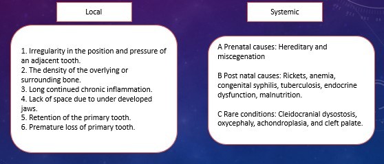
What impacts the teeth?
- Differences between the size of the teeth and the arch.
- The distal and mesial roots grow differently.
- Third molar delayed maturation: the tooth's dental development comes later than the skeleton's growth and development.
- There is less space available for the eruption of third molars during the mixed dentition era due to a decrease in the incidence of permanent molar extraction.
- Failure to develop the jaw bones as a result of eating more refined food results in insufficient functional stimulation for the development of the jaw bones.
Theories of impaction
Phylogenetic theory: Due to evolution, the human jaw size is becoming smaller and since the third molar tooth is the last to erupt there may not be room for it to emerge in the oral cavity.
Mendelian’s theory: Here genetic variation plays a major role if the individual genetically receives a small jaw from one parent and large teeth from another parent.
Nodine theory: The modern diet (soft diet) does not require a decided effort in mastication and hence, according to Nodine et al. the growth stimulus of the jaw is lost and modern man has impacted teeth.
Indications for extraction of impacted tooth
- Pericoronitis & peri coronal abscess
- Dental caries
- Periodontal dispoders
- Orthodontic causes
- Odontogenic cysts and tumours
- Management of unusual pain
- Resorption of the root of an adjoining tooth
- Teeth covered by a dental prosthesis
- Jaw fracture prevention
- Inflammation of the deep fascial space
- Before administration of radiotherapy
Contraindications
- Poor health status of the patient.
- The patient's advanced age.
- Damage to adjacent structures.
- The future status of the second molar is unclear.
- Third molars that are deeply affected but do not exhibit any relevant systemic or local pathology should not be extracted from patients.
Classification
Winter's classification
The Winter's classification is based on the inclination of the impacted wisdom tooth (3rd molar) to the long axis of the 2nd molar.
- Mesio-Angular
- Disto-Angular
- Horizontal
- Vertical
- Buccal / Lingual Obliquity
- Transverse
- Inverse
Extraoral examination
- The face and neck are examined for indications of infection, such as oedema or cheek redness.
- Anaesthesia and paresthesia are assessed on the lower lip.
- The regional lymph nodes are palpated for swelling and tenderness.
Intraoral examination
- Mouth opening.
- Oral cavity general examination: teeth, oral mucosa, oral hygiene.
- Area of the third molar.
- The state of the impacted tooth.
- First and second molar conditions.
- The amount of room that exists between the ascending ramus and the second molar's distal surface.
- Neighbouring bone.
- A fracture could make extracting an impacted third molar more difficult.
- It is important to recognise the pathological consequences of skeletal illnesses.
- Existence of tumours and cysts: Concerning an impacted tooth, both large and small eruption cysts may develop.
Radiography of impacted wisdom tooth
It is essential to get the intraoral and extraoral radiographs listed below:
- Periapical radiograph
- Occlusal X- ray of mandible
- Lateral oblique view of mandible
- Orthopantomogram
Surgical management
The steps in surgical removal of the third molar are as follows:
- Description of surgical access
- Analgesia
- The mucoperiosteal flap and incision
- Bone removal
- Removing the teeth
- Wound debridement
- Managing the bleeding
- Closure of wounds
- Follow-up post procedure

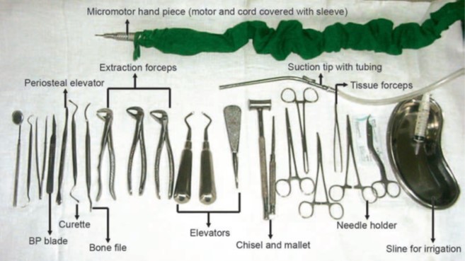
Armamenterium

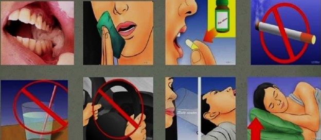
Post-operative instructions
Complications
Intra-operative
1. During incision
- Injury to the facial artery
- Injury to the lingual nerve
- Hemorrhage: A careful history should be taken
2. During bone removal
- Damage to the second molar
- Bur slipping into soft tissue and inflicting damage
- Extraoral/ mucosal burns
- Mandibular fracture caused by chisel and mallet use
- Subcutaneous emphysema
3. During elevation or tooth removal
- Luxation of neighbouring tooth/ fractured restoration
- Soft tissue injury due to slipping in the elevator
- Injury to the inferior alveolar neurovascular bundle
- Fracture of mandible
- Pushing a tooth root into the inferior alveolar nerve canal or submandibular space
- Breakage of instruments
- TMJ Dislocation: careful history mandatory
Post-operative Complications
1. Immediate
- Hemorrhage
- Pain
- Oedema
- Drug reaction
2. Delayed
- Alveolitis
- Infection
Disclaimer- The views and opinions expressed in this article are those of the author and do not necessarily reflect the official policy or position of M3 India.
About the author of this article: Dr Dhyey Saradhara is a practising dentist and an oral and maxillofacial surgeon from Surat.
-
Exclusive Write-ups & Webinars by KOLs
-
Daily Quiz by specialty
-
Paid Market Research Surveys
-
Case discussions, News & Journals' summaries