STEMI Localising Culprit Artery on ECG
M3 India Newsdesk Apr 21, 2023
ECG helps in viewing the electrical health of the heart and in cases of MI, it can predict which artery is occluded and the area at risk based on the ST changes in certain leads. This article may aid in the diagnosis and management of the patient.
Anterior MI
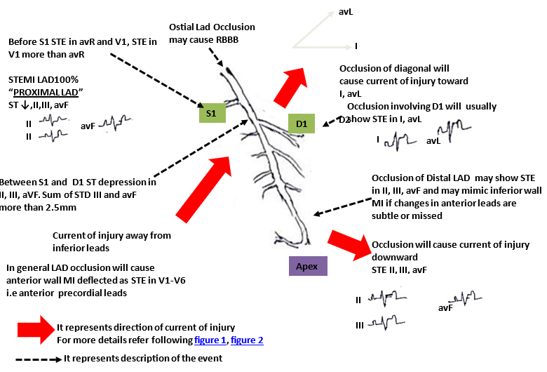
Details about the current of injury
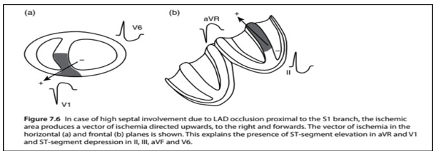
Figure 1: Current of if injury if occlusion involves first septal (S1)
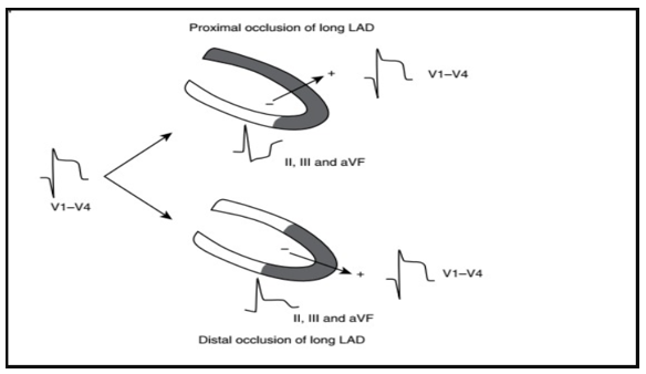
Figure 2: Current of injury in proximal vs Distal LAD and resulting different changes in ECG
Inferior MI
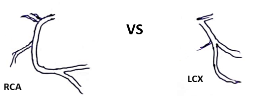
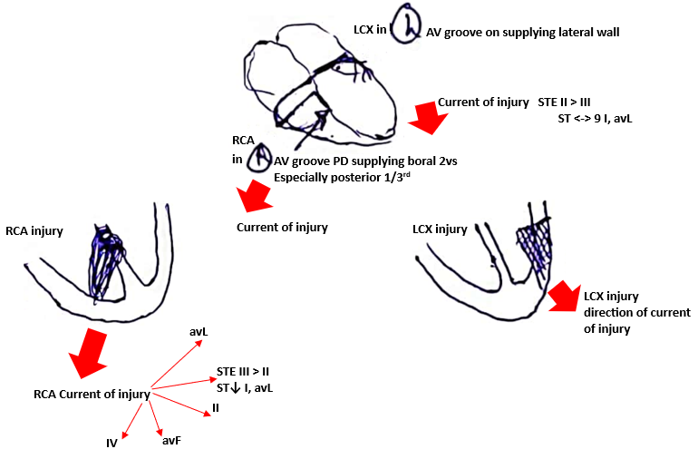
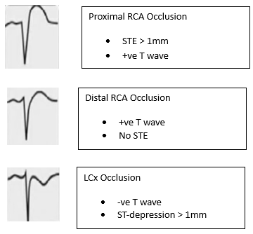
Lead V4R
Leads V3R and V4R should be recorded as rapidly as possible after the onset of chest pain because ST elevation persists for a much shorter period than STQ in extremity leads (II, III, and avF).
V3 to III ratio magnitude of ST depression in lead V3 relative to STE in lead III

ECG pattern STEMI equivalent

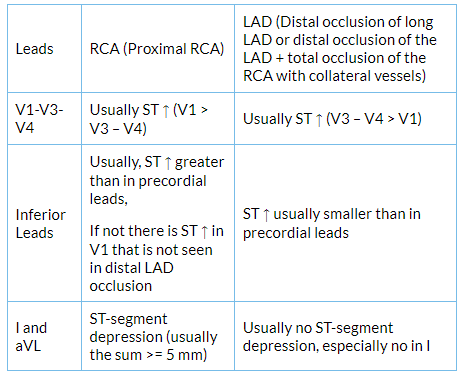
Differentiating pseudo AWMI from AWMI
Concomitant ST-segment elevation in the precordial leads (especially V1-V3-V4) and inferior leads (II, III and aVF).
Disclaimer- The views and opinions expressed in this article are those of the author and do not necessarily reflect the official policy or position of M3 India.
About the author of this article: Dr. Birdevinder Singh DM (Cardiology), Senior Consultant Interventional Cardiologist, Patiala Heart Institute, Patiala.
-
Exclusive Write-ups & Webinars by KOLs
-
Daily Quiz by specialty
-
Paid Market Research Surveys
-
Case discussions, News & Journals' summaries
