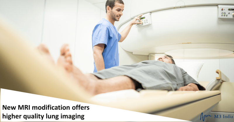
New MRI modification offers higher quality lung imaging
M3 Global Newsdesk Oct 30, 2019
Researchers at the NIH and Siemens have developed a new high-performance, low-field-strength MRI system that offers enhanced quality imaging of internal structures of the human body—particularly the heart and lungs—for improved disease diagnosis and treatment.

The results of their study, funded by the National Heart, Lung, and Blood Institute (NHLBI) division of the NIH, are published in Radiology.
“MRI of the lung is notoriously difficult and has been off-limits for years because air causes distortion in MRI images,” said lead author Adrienne E. Campbell-Washburn, PhD, Cardiovascular Branch, Division of Intramural Research, NHLBI, Bethesda, MD. “A low-field MRI system equipped with contemporary imaging technology allows us to see the lungs very clearly. Plus, we can use inhaled oxygen as a contrast agent. This lets us study the structure and the function of the lungs much better.”
Clinical MRI imaging in the United States is usually performed at 1.5 T or 3.0 T, but can go as high as 7.0 T for certain types of research. Of note, T is the SI unit Tesla, which measures magnetic flux density. Although high-field-strength MRI offers increased signal-to-noise ratio and resolution due to higher polarization, it also results in image distortion, higher specific absorption rates, restricted imaging efficiency, and higher costs.
“For some applications, low field strength may offer intrinsic advantages. At low field strength, short T1 and long T2* allow more efficient pulse sequence design; imaging near air-tissue interfaces is improved by virtue of reduced susceptibility gradients; and specific absorption rate is reduced, which can diminish heating of conductive devices and implants, and can eliminate pulse sequence parameter constraints,” wrote Dr. Campbell-Washburn and colleagues.
To that end, Dr. Campbell-Washburn and fellow researchers bucked the trend of using a high-field-strength MRI system by developing a high-performance, low-field-strength MRI system that allows for efficient imaging, MRI-guided catheterizations using metal devices, and MRI in sensitive regions. In addition to being safer for those with pacemakers and defibrillators, the new imaging method is also cheaper, quieter, and easier to install and maintain.
To create the 0.55-T MRI system, the researchers modified Siemens Healthcare’s Magnetom Aera, a 1.5-T commercial MRI system. Hardware and software required for high-quality images was kept intact. To assess the applications of the new system, Dr. Campbell-Washburn and colleagues performed 0.55-T MRI exams in 68 healthy participants and 15 participants with disease (average age: 34 years). Compared with images obtained at 1.5 T, the researchers were more clearly able to delineate lung cysts and surrounding tissue in patients with lymphangioleiomyomatosis at 0.55 T.
With respect to relaxation parameters, on average, T1 was 32% shorter, T2 was 26% longer, and T2* was 40% longer at 0.55 T compared with 1.5 T. Furthermore, the researchers noted that nine interventional devices made of metal were safe at 0.55 T (< 1 °C heating), with seven participants receiving MRI-guided right heart catheterization using metallic guidewires. Moreover, decreased image distortion was observed in the lungs, cranial sinuses, upper airway, and intestines due to enhanced field homogeneity, compared with 1.5 T. Inhalation of oxygen yielded lung signal enhancement of 19% at 0.55 T vs that of 7.6% at 1.5 T. Images of the brain, spine, heart, and abdomen were of good quality using 0.55 T.
“We can start thinking about doing more complex procedures under MRI-guidance now that we can combine standard devices with good quality cardiac imaging,” stated Dr. Campbell-Washburn. She also expressed that, in addition to the new technology’s clinical utility in cardiac and lung imaging, the system could aid with improved imaging of the brain, abdomen, and spine, and may offer greater clinical insight into speech and sleep disorders.
This story is contributed by Naveed Saleh and is a part of our Global Content Initiative, where we feature selected stories from our Global network which we believe would be most useful and informative to our doctor members.
-
Exclusive Write-ups & Webinars by KOLs
-
Daily Quiz by specialty
-
Paid Market Research Surveys
-
Case discussions, News & Journals' summaries